- Visibility 64 Views
- Downloads 16 Downloads
- Permissions
- DOI 10.18231/j.achr.2020.057
-
CrossMark

Abstract
Squamous cell carcinoma of kidney is the extremely rare entity of upper urinary tract neoplasm. The most common type of kidney cancer is clear cell renal cell carcinoma followed by papillary and chromophobe renal cell carcinoma. We report a case of renal cell carcinoma who presented with complaints of pain abdomen and hematuria. Radiologic investigation showed a renal mass and calculi in the left kidney. The patient subsequently underwent left radical nephrectomy. Pathological diagnosis of SCC of kidney was made. We report a rare case of primary renal squamous cell carcinoma involving whole of the kidney. With the best of our knowledge, only few cases of primary renal squamous cell carcinoma are reported.
Introduction
Cancer is one of the major causes of mortality in the world.[1] Kidney cancer is considered as the most deadly cancer of the urinary tract.[2] It is the ninth and fourteenth common cancer cases in men and women, respectively. Also, it is the sixteenth cause of death from cancer in the world.[3] Squamous cell carcinoma (SCC) of the kidney is an extremely rare entity, constitutes less than 1% of all urinary tract neoplasms.[4] and is mostly associated with long standing renal stone disease. This tumor has a poor prognosis because of its aggressive nature.[5]
Case Report
A 67-year-old female patient presented with complaints of pain abdomen and hematuria from last two weeks. Ultrasound examination revealed left kidney mass with nephrolithiasis. CT scan of abdomen showed large lobulated enhancing partially exophytic necrotic mass involving the upper-mid pole of left kidney favours the possibility of exophytic left renal cell carcinoma. The patient was operated in the surgery department and nephrectomy specimen was send to our department of pathology, GGS Medical College, Faridkot.
Gross Examination
The left kidney was found to be enlarged in size, measuring 12 cm × 7 cm × 6 cm. Cut section showed whole of the renal parenchyma replaced by grey white necrotic growth along with cystic areas filled with mucoid material and multiple yellow-brown colored stones ([Figure 1]). Grossly the growth was found involving the capsule. Ureter identified measuring 3cm in length. Cut section of perinephric fat showed focal grey white areas.

Microscopic examination
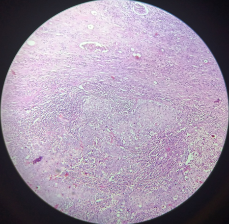
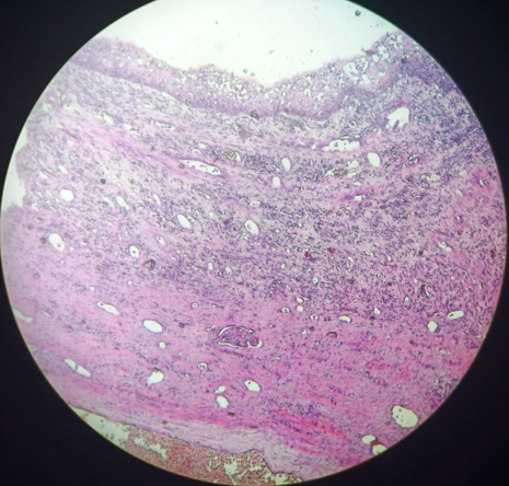
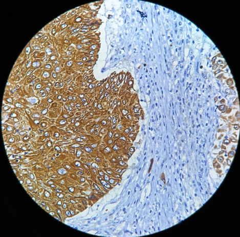
Hematoxylin and eosin stained tissue sections showed a cellular tumor with tumor cells arranged in nests. Individual tumor cells have round to oval nuclei, vesicular chromatin, prominent nucleoli and moderate amount of eosinophilic cytoplasm ([Figure 2]). The adjacent urothelium of pelvicalyceal system showed metaplasia and is replaced by stratified squamous lining ([Figure 3]). On immunohistochemistry by AEI/AE3 shows strong cytoplasmic staining of tumor cells ([Figure 4]). Pathological diagnosis of Moderately-Differentiated Squamous Cell Carcinoma of kidney was given. Tumor mass was involving the renal capsule and perinephric fat. Ureter and renal vessels were free of tumor.
Discussion
Primary renal squamous cell carcinoma is an extremely rare entity. Most squamous cell carcinoma of the kidney are moderately or poorly differentiated. Squamous cell carcinoma of kidney are more invasive than transitional cell carcinomas at diagnosis.[5], [6] The patients usually presented with advanced stage, making surgical resection difficult. This tumor has poor response to surgery, radiotherapy and chemotherapy resulted in a poor prognosis and short survival.[6], [7] Renal squamous cell carcinoma is mostly associated with chronic infections, renal calculi, radiotherapy or any factor that can irritate the urothelium. Normally the urothelium has transitional lining. Due to chronic irritation by stones it undergoes squamous metaplasia of lining epithelium which leads to dysplasia and become carcinogenic.[8], [9] In our case chronic irritation occurs by stones which leads to squamous metaplasia of urothelial lining and develops into cancer.
Previous studies in which squamous cell carcinoma of kidney is documented are
|
Study |
Laterality |
Site |
Nephrolithiasis |
|
Hipparagi SB et al in 2016 [10] |
Right |
Middle and Lower pole |
Present |
|
TK Sahoo et al in 2015 [11] |
Right |
Upper Pole |
Absent |
|
Ghosh P et al in 2014 [12] |
Right |
Lower Pole |
Absent |
|
Salehipour M et al in 2019 [13] |
Right |
Whole Kidney |
Present |
|
Lunney A et al in 2019 [14] |
Right |
Upper Pole |
Present |
|
Our Study |
Left |
Whole Kidney |
Present |
[Table 1] Shows:
Right kidney is most frequently affected in studies conducted by Hipparagi et al, TK Sahoo et al, Ghosh P et al, Salehipour M et al and Lunney A et al. But in our study Left kidney is involved by tumor.
Lower pole is involved in studies conducted by Hipparagi and Ghosh and upper pole is involved in studied conducted by TK Sahoo et al and Lunney A et al. Our study is in concordance with Salehipour M et al involving the whole kidney.
Nephrolithiasis is mostly associated in these studies, our case also shows association with nephrolithiasis.
Imaging techniques can show hydronephrosis, stones, and solid mass, but these findings are not specific for renal squamous cell carcinoma. Therefore, histopathological studies are very important for diagnosis. As compared to previous case reports, very little has been documented about squamous cell carcinoma of whole renal parenchyma. In our case whole kidney is involved by the tumor and is associated with nephrolithiasis.
Conclusion
Primary renal squamous cell carcinoma is an extremey rare tumor of upper urinary tract. It shows a strong association with nephrolithiasis. Imaging features on renal squamous cell carcinoma are non-specific. Histopathological findings are very important in the diagnosis of renal squamous cell carcinoma.
Source of Funding
None.
Conflicts of Interest
None
References
- Arabsalmani M, Mirzaei M, Ghoncheh M, Soroush A, Towhidi F, Salehiniya H. Incidence and mortality of liver cancer and their relationship with the human development index in the world. Biomed Res Ther. 2016;3(9):800-7. [Google Scholar]
- Mohammadian M, Pakzad R, Towhidi F, Makhsosi BR, Ahmadi A, Salehiniya H. Incidence and mortality of kidney cancer and its relationship with HDI (Human Development Index) in the world in 2012. Med Pharm Rep. 2017;90(3):286-93. [Google Scholar]
- Ferlay J, Soerjomataram I, Dikshit R, Eser S, Mathers C, Rebelo M. Cancer incidence and mortality worldwide: Sources, methods and major patterns in GLOBOCAN 2012. Int J Cancer. 2015;136(5):E359-86. [Google Scholar]
- Jain A, Mittal D, Jindal A, Solanki R, Khatri S, Parikh A. Incidentally Detected Squamous Cell Carcinoma of Renal Pelvis in Patients with Staghorn Calculi: Case Series with Review of the Literature. ISRN Oncol. 2011;2011:1-6. [Google Scholar]
- Mardi K, Kaushal V, Sharma V. Rare coexistence of keratinizing squamous cell carcinoma with xanthogranulomatous pyelonephritis in the same kidney: Report of two cases. J Cancer Res Ther. 2010;6(3):339-41. [Google Scholar]
- Lee T, Ko S, Wan Y, Cheng Y, Yang B, Huang D. Renal squamous cell carcinoma: CT findings and clinical significance. Abdominal Imaging. 1998;23(2):203-8. [Google Scholar]
- Berz D, Rizack T, Weitzen S, Mega A, Renzulli J, Colvin G. Survival of Patients with Squamous Cell Malignancies of the Upper Urinary Tract. Clin Med Insights: Oncol. 2012;6:11-8. [Google Scholar]
- Li MK, Cheung WL. Squamous cell carcinoma of the renal pelvis. J Urol. 1987;138:269-71. [Google Scholar]
- Holmäng S, Lele SM, Johansson SL. Squamous Cell Carcinoma of the Renal Pelvis and Ureter: Incidence, Symptoms, Treatment and Outcome. J Urol. 2007;178(1):51-6. [Google Scholar]
- Lunney A, Warraich I, Sharma P. Incidental Primary Squamous Cell Carcinoma of the Kidney Within a Calyceal Diverticulum Associated With Nephrolithiasis. Kidney. 2019;33(2). [Google Scholar]
- Ghosh P, Saha K. Primary intraparenchymal squamous cell carcinoma of the kidney: a rare and unique entity. Case Rep Pathol. 2014. [Google Scholar]
- Sahoo TK, Das SK, Mishra C, Dhal I, Nayak R, Panda AI. Squamous cell carcinoma of kidney and its prognosis: a case report and review of the literature. Case Rep Urol. 2015. [Google Scholar]
- Hippargi SB. Urothelial and Squamous Cell Carcinoma of Renal Pelvis – A Rare Case Report. J Clin Diagn Res. 2016;10(9). [Google Scholar]
- Salehipour M, Dastgheib N, Hosseinzadeh M, Makarem A, Rezvani A, Sanati A. Primary renal pelvis and ureter squamous cell carcinoma (SCC): a rare case report and review of literature. Int Med Case Rep J. 2019;Volume 12:189-92. [Google Scholar]