- Visibility 507 Views
- Downloads 129 Downloads
- Permissions
- DOI 10.18231/j.achr.2024.030
-
CrossMark

Epidermal inclusion cyst of external auditory canal– A common lesion at a rare site diagnosed by FNAC
Abstract
Epidermal inclusion cyst (EIC) is a benign condition that occurs rarely in the External auditory canal. EIC refers to cysts that result from the proliferation and implantation of epidermal elements within a circumscribed space in the dermis. Epidermal cysts occur anywhere in the body and are more common on the face, trunk, neck, extremities and scalp. The occurrence of EIC in the External auditory canal is rare. Lesions of such nature are frequently thought to be lesions like polyp, haematoma, haemangioma and are not included as one of the main differential diagnoses of lesions in External auditory canal. We report case of EIC of the External auditory canal in 4 years old child so that it should be kept as differential diagnosis of lumps in external auditory canal. The diagnosis was made on FNAC which is simple, OPD procedure and does not require any anaesthesia underlining the importance of FNAC in Ear lesions.
Introduction
Epidermal cyst is a benign keratin filled cyst of pilosebaceous origin. This can occur at any site in the body with common sites being head and neck region, trunk and extremities. An epidermal inclusion cyst refers to a cyst that results from the proliferation and implantation of epidermal elements within a circumscribed space in the dermis. These cysts grow through accumulation of epithelial and keratinous debris. Histopathologically, these lesions are formed by inclusion of keratinizing squamous epithelium within the dermis resulting in a cyst filled with lamellated keratin. Epidermal cyst are rare in External auditory canal and mostly presents as a palpable lump varying from 2 mm to 2.0 cm. The aim of this case report is to consider epidermal cyst at rare location of external auditory canal, do a clinical and cytopathological correlation and evaluate the role of Fine Needle Aspiration Cytology (FNAC) in its diagnosis.
Case Report
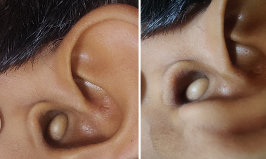
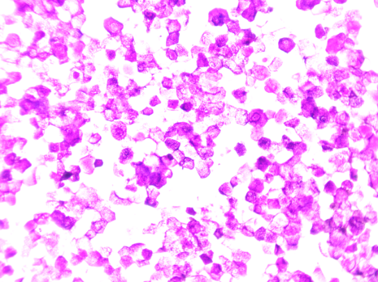
A 4 year old male child presented to ENT OPD with complaint of small lump in the left External auditory canal for one year which gradually started increasing in size. On examination 6×4 mm, non fluctuant, soft to firm, non tender lump was present in the External auditory canal.([Figure 1]) There was no pain or hearing loss associated with the lump. The patient had history of infection in the ear about one year back The patient was referred for FNAC to confirm diagnosis. FNAC was performed using 21 G needle with all aseptic precautions. FNA yielded few drops of fluid with pultaceous material. The cyst reduced in size to very minimal size. FNA smears were stained with giemsa stain and smears shows numerous anucleate squames and nucleate benign squamous cells. There were no atypical cells. Diagnosis was given as Epidermal Inclusion Cyst.([Figure 2]). FNAC diagnosis is very helpful in case of epidermal Inclusion cyst. As swelling subsided after FNAC biopsy or excision was not done.
Discussion
Epidermoid/epidermal inclusion cysts are cystic malformations lined with squamous epithelium. In the head and neck region, the most common location is the lateral eyebrow. Head and neck account for about 1.6–7.0%. Sublingual, submental and submandibular spaces are common localization in the floor of mouth. The head and neck sites affected most frequently with cutaneous cysts were the scalp (34%), neck (18%), periorbital area (17%), cheeks & lips (16%), periauricular area (9%), and nasal area (including forehead, 6%).[1]
Ihshan A et al presented a rare case of epidermal cyst of the bony external auditory canal (EAC) in an adult. Epidermal cyst of the bony EAC, although very rare, should be kept in the list of differential diagnosis of a skin-lined mass of the EAC. [2] Cho Y and lee DH analysed 15 cases of idiopathic epidermoid and dermoid cysts. Of 15 cases, 14 cases were epidermoid cysts. This study did not show any site preference for occurrence; the location of epidermoid cysts was evenly distributed at the ear canal, at the preauricular area, at the lobule, and at the postauricular area. [3] Alobaid et al reported bilateral epidermal cyst of external auditory canal in 28 years old women who presented with bilateral otalgia. On examination there was small cysts on both sides which was clinically suspected and confirmed as epidermal cyst on biopsy. [4] Abdel Aziz reported 9 paediatric patients of epidermoid cysts of the EAC and suggested Epidermoid cyst should be listed in the differential diagnosis of EAC masses. [5] Yalcin A et al reported Epidermal inclusion cyst of External auditory canal in 23 year old woman who had otitis for 2 months. [6] Bakshi JB, Subhash A reported Epidermoid cyst of external auditory canal in 12 month old child. Child had no hearing impairment. It presented as canal atresia.[7] Kim GW et al studied 17 cases of Epidermoid cysts of external auditory canal during period from 2004 to 2013. They studied their clinical presentation, size, ear symptoms, hearing problems. CT scan was done to see the extent before surgery. All cases were confirmed as Epidermoid cysts histo pathologically. Out of 17 cases 8 were bony EAC and 9 were cartilaginous EAC. Swelling and otalgia were common symptoms but 47 % of cysts were found incidentally. The size of Epidermoid cysts were 6.7 mm (range varied 2- 20 mm). [8] Taneja A et al reported EIC on FNAC at pinna in 4 year old child as rare case. [9] FNAC is confirmatory in the presence of a typical pultaceous aspirate and cytomorphological features of EIC. Therfore, FNAC plays a crucial role in its diagnosis and management.
Conclusion
In conclusion, we should consider EIC as differential diagnosis in case of any soft, cysic lump in External auditory canal. FNAC plays a very important role in providing accurate diagnosis of EIC.
Source of Funding
None.
Conflict of Interest
None.
References
- Sunil S, Oomnen N, Rathy R, Rekha V, Divya R, Sruthy V. Sruthy VK Epidermoid cyst of head and neck region. Int J Odontostomat. 2014;8(2):165-9. [Google Scholar]
- Ihshan A, Rauf A, Irfan I, Imtiaz D. Epidermal cyst of the bony external auditory canal. Indian J Otology. 2012;18(1):28-9. [Google Scholar]
- Cho Y, Lee D. Clinical characteristics of Idiopathic Epidermoid and dermoid cysts of ear. J Audiol Otol. 2017;21(2):77-80. [Google Scholar]
- Ahmad A, Jalagandeesh B. Bilateral Epidermoid Cyst of the External Auditory Canal: A Rare Case Report. Saudi J Otorhinolaryngol Head Neck Surg. 2023;25(3):131-4. [Google Scholar]
- Abdel-Aziz M. Epidermoid cyst of the external auditory canal in children: diagnosis and management. J Craniofac Surg. 2011;22(4):1398-400. [Google Scholar]
- Yalcin A, Hasan M, Ermin K, Buge O. Epidermal inclusion cyst of External auditory canal. J Craniofac Surg. 2010;21(4):1290-1. [Google Scholar]
- Bakshi J, Subhash A. Epidermoid cyst of external auditory canal presenting as canal atresia- A rare case report. Otolaryngol Head Neck Surg. 2016;2(1):106-7. [Google Scholar]
- Kim G, Park J, Kwon O, Kim D. Kim CW Clinical characteristics of Epidermoid cysts of external auditory canal. J Audiol Otol. 2016;20(1):36-40. [Google Scholar]
- Taneja A, Kaur P, Singh P, Bali I. Forget me not: Epidermal inclusion cyst of ear pinna, A common lesion at unusual location- a case report. Indian J Med Pharm Res. 2023;4(2):101-4. [Google Scholar]