Introduction
During pregnancy there is progressive anatomical, physiological and biochemical changes not only confined to the genital organs but also to all systems of the body.1 any of these changes begin soon after remarkable adaptations occur in response to physiological stimuli provided by the fetus or fetal tissues. Equally astounding is the fact that the woman who is pregnant returns almost completely to her pre-pregnancy state after delivery and cessation of lactation.2 The understanding of these adaptations to pregnancy is necessary. Because, for example hemoglobin concentration less than 12 gm /dl which define anemia in general population, is not considered indicative of anemia in pregnancy because of a physiological change called hemodilution of pregnancy.
According to WHO, Anemia in pregnancy is defined as a hemoglobin concentration of less than 110 g/L (less than 11 g/dL) in venous blood.3 Anemia is the most common nutritional deficiency disorder in the world. WHO has estimated that prevalence of anemia in developed and developing countries in pregnant women is 14 per cent in developed and 51 per cent in developing countries and 65-75 percent in India.4
Studies carried out in India and elsewhere have shown that iron deficiency is the major cause of anemia followed by folate deficiency. Pregnant women are at higher risk due to their increased need. It is almost universally accepted that hematological parameters like Hb, HCT, MCV, MCH, MCHC, RDW as well as peripheral smear examination are important diagnostic tools for diagnosing anemia in pregnant women.
Materials and Methods
In the present study, 1000 pregnant women who attended antenatal outpatient department of one of the government teaching hospitals of Gujarat were included. Detailed history including age, socio-economic status, gap between two pregnancies, gravid status and trimester status was taken. After taking detailed history and written consent a blood sample was taken in EDTA vacuette. Blood samples from 1000 antenatal women who attended antenatal outpatient department were collected for evaluation of various hematological parameters. After collecting the blood it was transferred into a properly labeled vacuette. This vacuette was used for analyzing various hematological parameters (Hb, RBC count, HCT, MCV, MCH, MCHC & RDW) with the help of automated hematology analyser. Also a blood smear was prepared from the same vacuette and it was study simultaneously and the P/S findings were noted.
Results
A total of 1000 patient samples were studied in this study. Various hematological parameters were noted by automated hematology analyzer and peripheral smear was also studied from each sample.
Table 1
Socio demographic and obstetric profile of study group
Women were mainly between the age of 20 and 25 years with youngest being 18 years and oldest being 40 years. Majority (85.2%) had education of above secondary school level. Two thirds (61.2%) of them were not working. Majority (93.1%) belonged to lower socioeconomic class as per modified Prasad classification. Out of 1000 women, 437 (43.7%) were second gravida, 347 were primigravidae, 187 were third gravida and 29 were fourth gravidae. (Table 1)
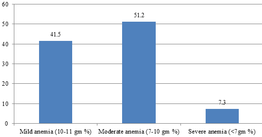
Total 537(55 %) women had hemoglobin less than 11 gm%, which means that they were anemic according to WHO guidelines. Around 7.3 % of females had severe anemia. (Figure 1)
Table 2
| Socioeconomic class | Hemoglobin value | Total | Chi square | P value | |
| <11 gm% | >=11 gm% | ||||
| Lower | 504 (54.1%) | 427(45.9%) | 931 | 1.02 | 0.31 |
| Middle | 33(47.8%) | 36(52.3%) | 69 | ||
Comparison of hemoglobin in women from lower and middle socioeconomic class
As seen in table 2, women with lower socioeconomic status have more prevalence of anemia as compared to women with middle socioeconomic status. The difference was not significant statistically by applying chi square test.(p<0.05)
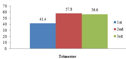
Figure 2 reveals that prevalence of anemic women increases from 1st trimester to third trimester.
Table 3
Various hematological parameters in women of different trimesters
Comparison of mean of various parameters in women in different trimesters is shown in Table 3. This comparison shows that women in 3rd trimester have less hemoglobin, RBC count and HCT as compared to others.
Table 4
Various peripheral smear findings in anemic women
Around 65.7 % women have hypochromic microcytic picture in peripheral smear which is probably due to iron deficiency in these women. Two women showed sickle like cells in the peripheral smear. (Table 4)
Discussion
Anemia in pregnant women is very common and has serious health implications. Severe anemia during pregnancy significantly contributes to maternal mortality and morbidity.5 There is evidence that severe anemia also increases perinatal morbidity and mortality by causing intrauterine growth retardation and preterm delivery. In India, the prevalence of anemia in pregnant women has been reported to be in the range of 33% to 89%. So with the aim of studying prevalence of various types of anemia in women presented to antenatal outpatient department, this study was carried out which included 1000 women.
Most common age group of pregnant women in present study is 20-25 years, which is comparable to Bisoi et al6 and Virender Gautam et al.7
In present study the prevalence of anemia was 53.6% which was comparable to Bentley and Griffith8 (48.8%), but it was considerably higher in Bisoi et al6 (67.8%), Virender Gautam et al7 (96.5%), Nadeem ahmad et al9 (74.8%) and Toteja et al10 (84.9%). However the findings in present study is quite nearer to National Family Health Survey 4 (NFHS- 4) conducted during 2015-16 which found an overall prevalence of 50.3% among pregnant women.11
In present study 41.5% women were having mild anemia, 51.2% women were having moderate anemia and 7.3% women were having severe anemia, which is somewhat comparable to Bisoi et al6 & Bentley and Griffith.8 In present study percentage of anemic primigravidae women was 33.9% and anemic multigravidae was 66.1% which is comparable to Debolina Sahoo et al.12
In present study percentage of anemic women in 1st, 2nd and 3rd trimester was 19.1%, 41.9% and 39% respectively which is comparable to Bisoi et al6 (18.3%, 61.6%, 20.1%). Debolina Sahoo et al12 shown little variation (25%, 32.5%, 42.5%).
In present study the hypochromic microcytic peripheral smear picture was seen in 65.7% women which is comparable to Chamakuri Nirmala et al (69.2%)13 and normochromic normocytic picture was seen in 37.2% women which is comparable to Erli Amel Ivan et al (39.3%).14
Conclusions
The present study comprises of 1000 antenatal women that presented to antenatal OPD at one of the medical colleges of Gujarat. The prevalence of anemia in pregnant women is still at a very high level in spite of the efforts from the government. Although they are supplied with iron folic acid tablets during the pregnancy, still the women showed low hemoglobin in third trimester, which shows that there is an inherent nutritional deficiency in pre-pregnancy period and also during pregnancy. Study of hematological parameters and peripheral smear can very well guide to classify the anemia in antenatal women so that women can be supplied with appropriate treatment and the fetomaternal outcome can be improved.


