- Visibility 73 Views
- Downloads 12 Downloads
- DOI 10.18231/j.achr.2021.053
-
CrossMark
- Citation
Application of IAC Yokohama system for breast cytology – The experience at a tertiary care hospital
- Author Details:
-
Aka Sunitha
-
Sharath Chandra
-
GVRN Krishna Kanth *
Introduction
Breast malignancies are one of the commonest malignancies in Indian women. Increasing urbanization is attributed to raise of breast cancers and have surpassed cervix cancer in recent times and are now ranked top in some metros. [1] FNA is used as an important diagnostic tool as a part of triple assessment. The international academy of cytology system for reporting breast fine needle aspiration cytology was formed in the year 2016 by a set of cytopathologists, radiologists, surgeons, and oncologists. [2] According to IAC Yokohama system of reporting breast cytology, 5 different categories have been classified as follows:
Insufficient / Inadequate
Benign
Atypical, probably benign
Suspicious of malignancy
Malignant
This type of reporting is helpful in organizing the way of reporting breast FNA samples. It provides more appropriate information to the clinician in decision making for patient care management and improves mutual communication.[3] Also the FNA procedure has the advantage of being easier, quicker to perform and less cumbersome. By using the IAC Yokohama system the samples received in our pathology department were classified into different categories and ROM was also calculated for the same.
Materials and Methods
The present study was carried out in the department of pathology, ESIC medical college and hospital, Sanathnagar, Hyderabad, India. The study was carried out over a period of 2 years, retrospectively from June 2019 to May 2021. 305 breast FNA samples were collected and categorized according to the IAC Yokohama system for reporting breast cytopathology. All were female patients. 95 cases of these had histopathological correlation and statistical parameters, ROM were calculated on the obtained data.
P-value was also calculated by using the chi-square test.
Results
A total of 305 FNA samples were obtained and all these are categorized according to the IAC Yokohama system after thorough review by an experienced pathologist. The age group for all the cases was 11 – 80 yrs ([Table 1]) and those cases with palpable breast lump were taken. The most common age group with lump in the breast observed in our study was 31 – 40 yrs. The earliest age presented with breast malignancy was 33 and we have observed 10 malignancies in the age group of 31 – 40 yrs.
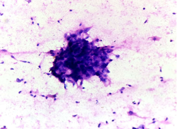
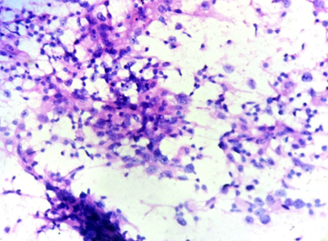
All the FNA samples obtained are put in one of the 5 categories according to the IAC Yokohama system of reporting. The percentage distribution of cases according to IAC Yokohama are shown in ([Table 2]).
For 305 FNA samples received, 95 cases were obtained for HPE correlation ([Table 3]). The ROM is calculated for each category. Sensitivity, Specificity, PPV, NPV, Diagnostic accuracy are calculated for the present study using MS EXCEL sheet.
Chi-square test for categorical data was used for calculation of p-value to assess statistical significance ([Table 4]). p-value < 0.05 is considered as significant. In the present study p- value obtained was 0.0001 which is very significant.
|
Age range |
No. of cases |
|
11 – 20 |
24 |
|
21 – 30 |
72 |
|
31 – 40 |
96 |
|
41 – 50 |
81 |
|
51 – 60 |
27 |
|
61 – 70 |
4 |
|
71 – 80 |
1 |
|
IAC yokohama category |
Total no. of cases |
% of distribution |
|
Insufficient |
21 |
6.89% |
|
Benign |
221 |
72.46% |
|
Atypical |
10 |
3.28% |
|
Suspicious of malignancy |
10 |
3.28% |
|
Malignant |
43 |
14.09% |
|
Total |
305 |
100% |
|
IAC yokohama category |
Cytology |
Histopathology |
|
Insufficient |
21 |
4 |
|
Benign |
221 |
50 |
|
Atypical |
10 |
5 |
|
Suspicious of malignancy |
10 |
7 |
|
Malignant |
43 |
29 |
|
Total |
305 |
95 |
|
IAC yokohama categories |
Benign |
Malignant |
Total |
Chi- square value |
P-value |
|
Insufficient |
4 |
0 |
4 |
83. 115 |
<0.0001 |
|
Benign |
49 |
1 |
50 |
||
|
Atypical |
5 |
0 |
5 |
||
|
Suspicious of malignancy |
2 |
5 |
7 |
||
|
Malignant |
0 |
29 |
29 |
||
|
Total |
58 |
35 |
95 |
|
IAC yokohama categories |
ROM |
|
Insufficient |
0% |
|
Benign |
2% |
|
Atypical probably benign |
0% |
|
Suspicious of malignancy |
71.43% |
|
Malignant |
100% |
|
Statistical parameters |
Percentage |
|
Sensitivity |
100% |
|
Specificity |
96.66% |
|
Positive predictive value |
94.59% |
|
Negative predictive value |
100% |
|
Diagnostic accuracy |
97.89% |
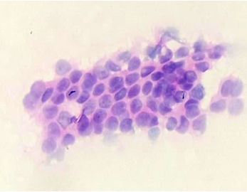
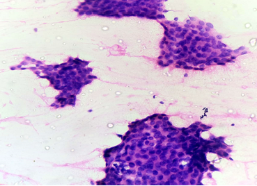
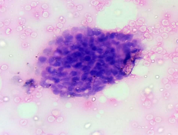
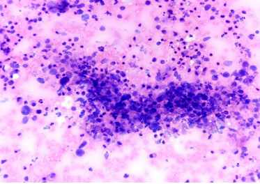
Discussion
Breast FNA is a quick, precise and highly convenient diagnostic procedure which can be done in OPD. It has less complications and can diagnose a wide spectrum of diseases with high accuracy and has less turnaround time than HPE. So in this era of core biopsies, FNA is still continuing as an important diagnostic tool along with clinical imaging and self assessment as a part of triple assessment. It has high sensitivity, specificity and high reproducibility.[4], [5], [6], [7], [8], [9], [10], [11]
In our study, we observed the most common age group affected with breast malignancies are between 31 – 40 yrs and the earliest age group presenting with breast lump was 13 years. The present study is comparable to Badge SA et al [12] and Sreedevi CH et al[13] where there are no malignant cases in the age group of 11-20yrs. In our study we have observed 1 malignant case in the age group 21-30 and no malignant cases between 71-80. This could be because of increased incidence of breast cancers in younger age group when compared to elderly probably because of urbanization. The % of benign and malignant cases obtained in the present study is comparable to the studies of Sreedevi CH et al[13] and Badge SA et al. [12]([Table 7])
|
Age |
Sreedevi CH et al [13] |
Badge SA et al [14] |
Present study |
|||
|
Benign |
Malignant |
Benign |
Malignant |
Benign |
Malignant |
|
|
11-20 |
22 |
0 |
10 |
0 |
24 |
0 |
|
21-30 |
36 |
0 |
36 |
0 |
71 |
1 |
|
31-40 |
17 |
4 |
82 |
17 |
84 |
12 |
|
41-50 |
8 |
4 |
27 |
29 |
61 |
20 |
|
51-60 |
2 |
5 |
7 |
4 |
11 |
16 |
|
61-70 |
0 |
1 |
0 |
6 |
2 |
2 |
|
71-80 |
1 |
0 |
0 |
2 |
1 |
0 |
|
Total |
86(86%) |
14(14%) |
162(73.63%) |
58(26.37%) |
254(83.27%) |
51(16.73%) |
In the present study we obtained 305 breast FNA samples of female patients and these were categorized into one of the 5 given categories according to the IAC Yokohama system and the number of cases obtained in each category was subsequently compared with other studies done in India and
elsewhere showing that the relative proportion of malignant cases are growing in the Indian context.([Table 8])
|
IAC Yokohama Category |
Montezuma D et al., [14] |
Wong S et al., [15] |
Poornima V Kamatar et al., [16] |
Apuroopa M et al., [17] |
Present study |
|
Insufficient |
209 (5.77%) |
301 (11%) |
22 (5%) |
39 (4.3%) |
21 (6.89%) |
|
Benign |
2660 (73.38%) |
1937 (72%) |
332 (71%) |
522(58%) |
221 (72.46%) |
|
Atypical probably benign |
498 (13.74%) |
117 (4.3%) |
7 (1%) |
160 (17.7) |
10 (3.28%) |
|
Suspicious |
57 (1.57%) |
59 (2.2%) |
8 (2%) |
63 (7.2%) |
10 (3.28%) |
|
Malignant |
201 (5.54%) |
278 (10%) |
101 (21%) |
116 (12.8%) |
43 (14.09%) |
|
Total |
3625 |
2696 |
470 |
900 |
305 |
ROM calculated for each category was comparable to the study of Poornima V Kamatar et al., [16] In the present study we observed 0% ROM in insufficient and atypical categories. Atypia associated with malignant lesions has been appropriately recognized and was put up in higher categories which is responsible for low ROM with atypical category. The ROM is comparatively less in the present study when compared to other studies. So, more diligence has to be paid in categorizing suspicious lesions.([Table 9])
|
IAC Yokohama Category |
Montezuma D et al, [14] |
Poornima V Kamatar et al., [16] |
Apuroopa M et al., [17] |
Present study |
|
Insufficient |
4.8% |
0% |
5.0% |
0% |
|
Benign |
1.4% |
4% |
1.2% |
2% |
|
Atypical probably benign |
13% |
66% |
12.5% |
0% |
|
Suspicious |
97.1% |
83% |
93.65% |
74.43 |
|
Malignant |
100% |
99% |
100% |
100% |
The sensitivity, specificity, positive predictive value, negative predictive value, diagnostic accuracy are calculated and these statistical parameters are comparable to studies of Moschetta M et al [18] We observed 100% sensitivity and negative predictive value in our studies.
p-value is also calculated for present study and it is <0.0001 which is very significant and it implies that the recognition of various Yokohama categories in ESIC setup has largely been successful and is comparable to the established standards of IAC Yokohama and this has helped the department in giving appropriate guidance to the surgeons in dealing with breast lesions.(table 10)
|
Statistical parameters |
Montezuma D et al., [14] |
Moschetta M et al., [18] |
Poornima V Kamatar et al., [16] |
Apuroopa M et al.,[17] |
Present study |
|
Sensitivity |
97.56% |
97% |
94.59% |
95.9%, |
100% |
|
Specificity |
100% |
94% |
98.9% |
97.89%, |
96.66% |
|
Positive predictive value |
100% |
91% |
98.59% |
96.79%, |
94.59% |
|
Negative predictive value |
98.62% |
98% |
95.74% |
97.64% |
100% |
|
Diagnostic accuracy |
99.11% |
95% |
96.97% |
91.5 % |
97.89% |
Conclusion
Application of IAC Yokohama system of reporting breast cytopathology helps in easy categorization of breast FNA samples and it improves the efficacy of cytopathologist. It also provides better clarity to the clinicians in the management of the patient and can reduce unnecessary surgeries.
Conflicts of Interest
The authors declare that there are no conflicts of interest regarding the publication of this paper.
Source of Funding
None.
References
- G Agarwal, P Ramakant. Breast cancer care in India: The current scenario and the challenges for the future. Breast Care ( Basel) 2008. [Google Scholar] [Crossref]
- A S Field, F Schmitt, P Vielh. IAC standardized reporting of breast fine-needle aspiration biopsy cytology. Acta Cytol 2017. [Google Scholar]
- Field. The International Academy of cytology Yokohama System for Reporting Breast FNA Cytopathology. Acta Cytologica 2019. [Google Scholar]
- Y H Yu, W Wei, J L Liu. Diagnostic value of fine-needle aspiration biopsy for breast mass: a systematic review and meta-analysis. BMC Cancer 2012. [Google Scholar]
- B S Ducatman, H H Wang, E Cibas, B Ducatman. Breast. Cytology: principles and clinical correlates, 4th Edn. 2014. [Google Scholar]
- J Dong, A Ly, R Arpin, Q Ahmed, E Brachtel. Breast fine needle aspiration continues to be relevant in a large academic medical center: experience from Massachusetts General Hospital. Breast Cancer Res Treat 2016. [Google Scholar]
- A S Field, AS Field, MR Zarka. Fine needle aspiration biopsy cytology of breast: a diagnostic approach based on pattern recognition. Practical Cytopathology: A Diagnostic Approach to Fine Needle Aspiration Biopsy’ 2017. [Google Scholar]
- M Wang, X He, Y Chang, G Sun, L Thabane. A sensitivity and specificity comparison of fine needle aspiration cytology and core needle biopsy in evaluation of suspicious breast lesions: a systematic review and meta-analysis. Breast 2017. [Google Scholar] [Crossref]
- R S Hoda, R N Arpin Iii, R V Gottumukkala, K S Hughes, A Ly, E F Brachtel. Diagnostic value of fine-needle aspiration in male breast lesions. Acta Cytol 2019. [Google Scholar] [Crossref]
- D Montezuma, D Malheiros, F Schmitt. Breast FNAB cytology using the newly proposed IAC Yokohama System for Reporting Breast Cytopathology: the experience of a single institution. Acta Cytol 2019. [Google Scholar] [Crossref]
- S Wong, M Rickard, P Earls, L Arnold, B Bako, A S Field. The IAC Yokohama System for reporting breast FNAB cytology: a single institutional retrospective study of the application of the system and the impact of ROSE. Acta Cytol 2019. [Google Scholar] [Crossref]
- S A Badge, A G Ovhal, K Azad, A T Meshram. Study of fine-needle aspiration cytology of breast lumps in rural area of Bastar district Chhattisgarh. Med J DY Patil Univ 2017. [Google Scholar] [Crossref]
- C H Sreedevi, K Pushpalatha. Correlative study of FNAC and histopathology for breast lesions. Trop J Path Micro 2016. [Google Scholar] [Crossref]
- D Montezuma, D Malheiros, F C Schmitt. Breast Fine needle aspiration biopsy [7] cytology using the newly proposed IAC Yokohama system for reporting breast cytopathology: The experience of a single institution. Acta Cytol 2019. [Google Scholar]
- M Apuroopa. Application of Yokohama System for Reporting Breast Fine Needle Aspiration Cytology in Correlation with Histopathological and Radiological Findings. Ann Pathol Lab Med 2020. [Google Scholar]
- P V Kamatar. Breast Fine needle Aspiration Biopsy Cytology Reporting using International Academy of Cytology Yokohama System Two Year Retrospective Study in Tertiary Care Centre in Southern India. National J Lab Med 2019. [Google Scholar]
- S Wong, M Rickard, P Earls, L Arnold, B Bako, A S Field. The International [8] Academy of Cytology Yokohama System for reporting breast fine needle aspiration biopsy cytopathology: A single institutional retrospective study of the application of the system categories and the impact of rapid onsite evaluation. Acta Cytol 2019. [Google Scholar]
- M Moschetta. Comparison between fine needle aspiration cytology (FNAC) [9] and core needle biopsy (CNB) in the diagnosis of breast lesions. G Chir- J Surg 2014. [Google Scholar]
